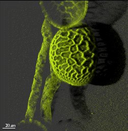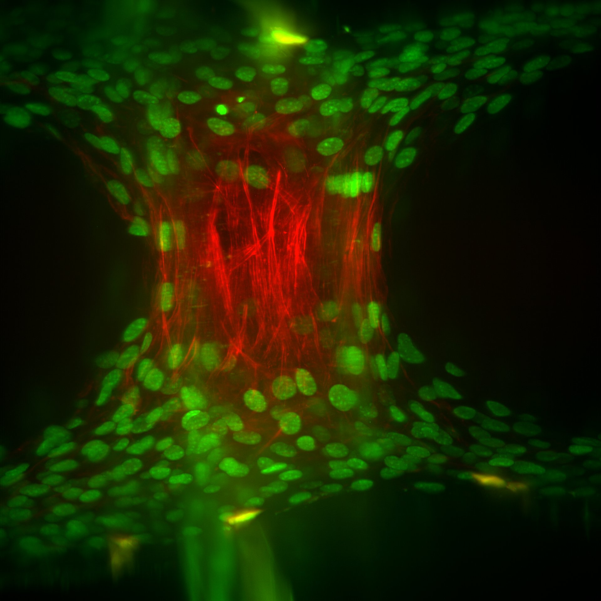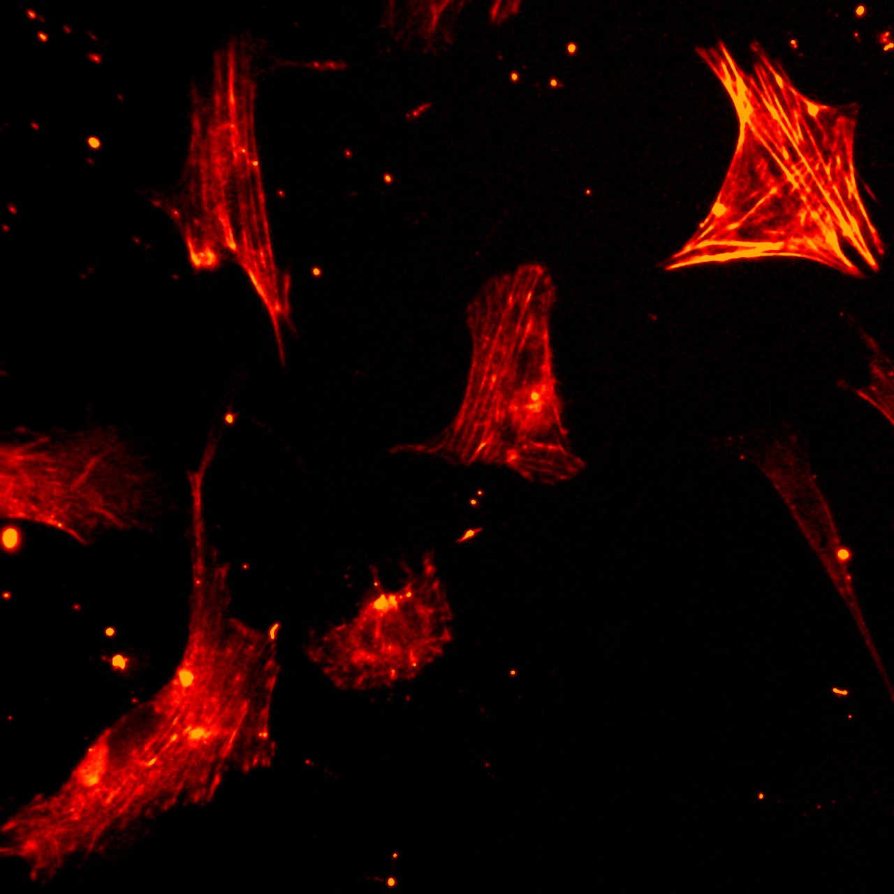
Modern fluorescence and light microscopical techniques provide powerful tools to investigate living organisms, tissues or cells. Live-cell imaging allows observing time-dependent changes in cells, tissue preparations or entire organoids. Using high resolution microscopy, e.g. STED (stimulated emission depletion), even structures below the optical resolution limit become visible, thus enabling new insights into the biology of cells and organs. Even the development of entire, small organisms may be observed using light sheet microscopy. In addtion, these techniques go beyond morphological or structural information but enable measuring important physiological parameters with genetically-engineered fluorescence indicators, e.g. pH, cytosolic Ca2+ or metabolite concentrations, in living cells.
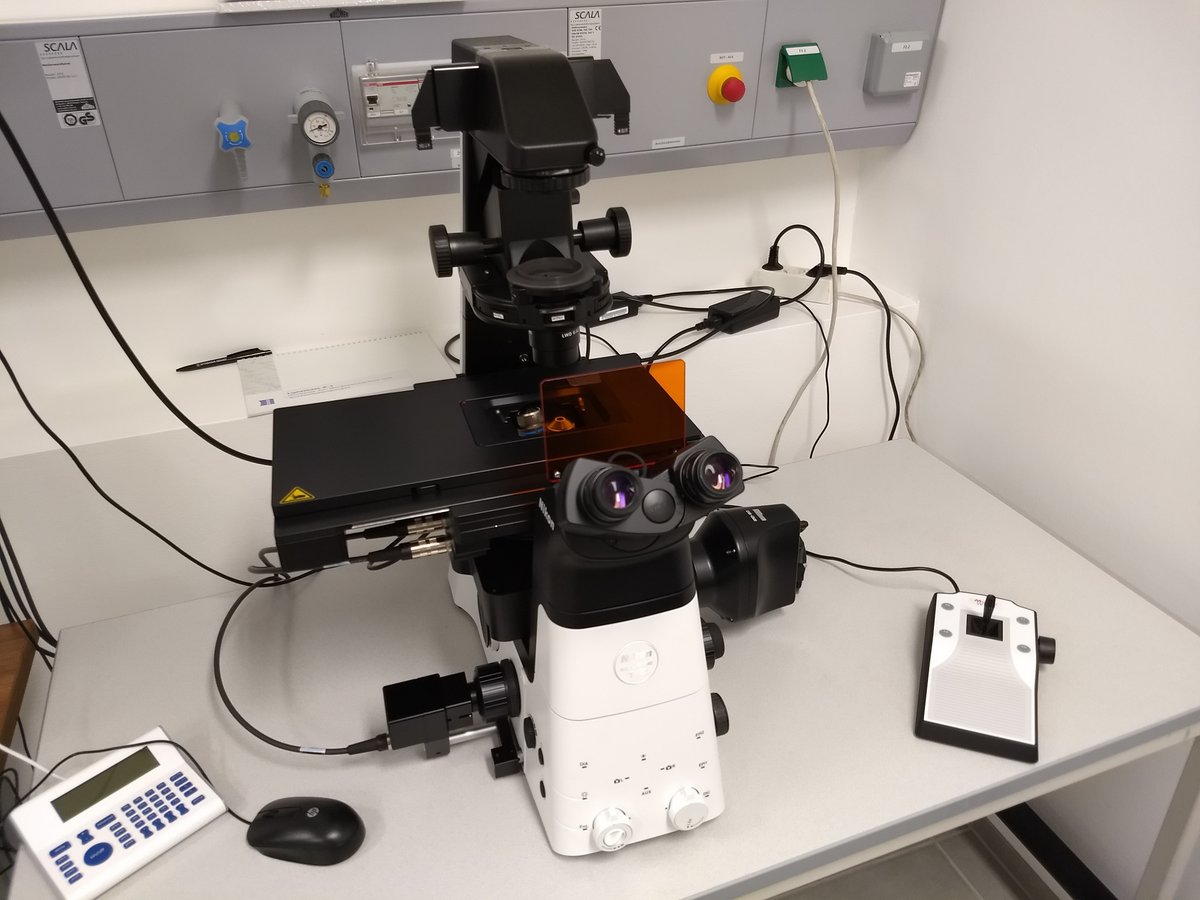
The Technology Platform Advanced Light and Fluorescence Imaging at the Paris-Lodron-University Salzburg offers all scientists access to fluorescence-based imaging techniques ranging from epi-fluorescence microscopy to super-resolution imaging and light sheet microscopy as well as analytical fluorescence microscopy. Support and help is given for first time users, advanced users may book the equipment using the core facility's webpage (see below).
7230 or 6205


You have to be logged in to download files.